Are you a dental or medical professional looking for a detailed guide to orthognathic surgery? “Introduction to Contemporary Orthognathic Surgery” is an essential resource that bridges the gap between theoretical knowledge and practical application in orthognathic procedures. As both a student and educator in maxillofacial surgery, I’ve found this book to be an invaluable companion for understanding the complexities of jaw surgery.
What Makes This Book Unique?

Unlike many technical surgical texts that focus solely on procedures, this book draws from the authors’ unique military surgical experiences to provide a comprehensive perspective on orthognathic surgery. From patient evaluation and treatment planning to surgical execution and complication management, the authors have created a workflow-oriented guide that’s accessible to both new residents and experienced surgeons.
Let’s explore what this book offers and why it deserves a place in every oral and maxillofacial surgeon’s library.
Who Are the Authors?
The book is authored by:
- Andrew C. Jenzer, DDS (MAJ, US Army)
- Jonathan L. Czerepak, DMD (formerly US Army)
- Joseph W. Ivory, DDS (LTC (R), US Army)
With contributions from numerous specialists, the book combines decades of clinical experience from both military and private practice settings.
Chapter-by-Chapter Summary
Chapter 1: The Evaluation Appointment
This foundational chapter provides a comprehensive framework for conducting thorough orthognathic evaluation appointments. Dr. Jenzer emphasizes that a precise and detailed workup is crucial for developing an accurate diagnosis and surgical plan.
Key Components of the Evaluation
The chapter outlines a systematic approach to patient evaluation:
- Patient History and Interview: Understanding the patient’s chief complaint, history of present illness, and previous orthodontic treatments is crucial. Dr. Jenzer emphasizes the importance of documenting any previous orthodontic therapy, as multiple rounds of orthodontics can lead to root resorption.
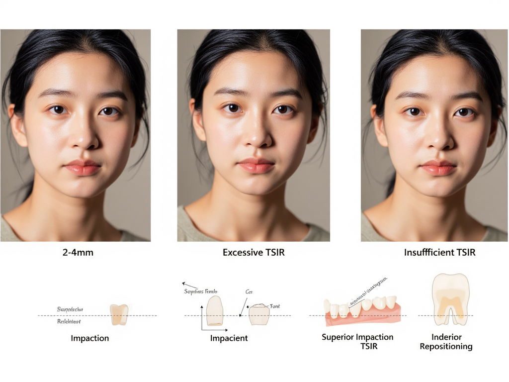
- Clinical Measurements: Critical measurements include tooth show in repose (TSIR), which is essential for determining the ideal position of the maxilla. The chapter demonstrates how to accurately measure TSIR by having the patient maintain their head in an upright position while breathing through their mouth.
- Midline Assessment: The midline alignment between maxillary and mandibular dentition is measured relative to the midsagittal plane, providing crucial information for symmetry correction.
- Cant Evaluation: Methods for measuring facial cant (sideways tipping of the jaw) are detailed, including techniques using a Fox plane and measuring from orthodontic brackets to the medial canthi.
- Photography Protocol: The chapter provides detailed instructions for standardized clinical photography, including extraoral (frontal repose, frontal animation, and lateral profile) and intraoral photographs. Lighting, positioning, and camera settings are discussed to ensure high-quality diagnostic images.
The author includes a sample orthognathic evaluation template as a practical tool that clinicians can customize for their practice, highlighting the importance of documenting specific measurements like TSIR, facial fifths, facial thirds, and midline discrepancies.
Chapter 2: The Presentation
This chapter focuses on synthesizing the clinical data into a comprehensive diagnostic presentation. Dr. Jenzer outlines how to analyze clinical photographs and radiographs systematically to develop a precise set of diagnoses.
Photographic Analysis
The chapter provides a detailed approach to analyzing extraoral and intraoral photographs:
- Frontal Repose Analysis: The author demonstrates how to evaluate facial fifths, brow position, nasal symmetry, TSIR, and midline alignment.
- Frontal Animation: Assessment includes Fitzpatrick and Glogau classifications, animation symmetry, tooth show percentage, and buccal corridor fill.
- Profile Analysis: Evaluation of facial profile type (concave, straight, or convex), facial thirds, radix assessment, nasal complex evaluation, nasolabial angle, labiomental fold, and neck contour using the Dedo classification.
- Intraoral Photography Analysis: The chapter covers midline relationships, dental abnormalities, evidence of bruxism, crossbite assessment, and arch form evaluation.
Radiographic Analysis
Dr. Jenzer provides guidance on interpreting radiographs:
- Panoramic Radiograph: Assessment includes evaluating condyle position, sinus conditions, missing teeth, and potential root resorption.
- Lateral Cephalometric Analysis: The chapter explains how to interpret and trace lateral cephalometric radiographs, including SNA, SNB, ANB angles, mandibular plane angle, and Wits appraisal. Dr. Jenzer emphasizes looking beyond numbers to understand the relationships between structures.
The chapter concludes with creating a comprehensive diagnosis list categorized into hard tissue, soft tissue, and dental diagnoses, which forms the foundation for treatment planning.
Chapter 3: Orthodontics and Treatment Planning
Dr. Russell M. Weaver provides the orthodontist’s perspective on orthognathic surgery, offering crucial insights into the interdisciplinary approach required for successful outcomes.
Understanding Orthodontic Terminology and Concepts
The chapter begins by familiarizing surgeons with orthodontic terminology:
- Tooth Numbering Systems: Explanation of the Palmer and alphanumeric systems used by orthodontists compared to the Universal numbering system.
- Cephalometric Analysis: Emphasis on using cephalometric values as directional guides rather than strict targets.
- Orthodontic Appliances: Detailed descriptions of brackets, bands, wires, ligation methods, and surgical hooks/lugs used in orthognathic cases.
Treatment Phases
Dr. Weaver divides orthognathic treatment into distinct phases:
- Pretreatment Evaluation and Coordination: The importance of orthodontist-surgeon communication in establishing treatment objectives for occlusion, stability, and esthetics.
- Presurgical Orthodontics: Detailed discussion of tooth extraction considerations, dental decompensation, arch coordination, preparation for segmentation, and creation of cut spaces for surgery.
- Surgery: Considerations for intraoperative coordination between orthodontist and surgeon.
- Postsurgical Orthodontics: Management of the patient after surgery, including splint removal, final occlusal settling, and retention protocols.
The chapter emphasizes the critical role of communication between the orthodontist and surgeon throughout all phases of treatment, particularly in planning dental decompensation and arch coordination.
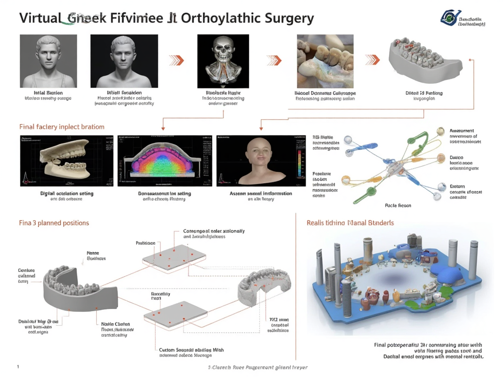
Chapter 4: The Process of Surgical Planning
This chapter, co-authored by Drs. de Latour and Jenzer, focuses on translating clinical data into a concrete surgical plan, with particular attention to virtual surgical planning (VSP).
Setting the Occlusion
The authors describe various methods for setting the final occlusion:
- Digital vs. Physical Methods: Comparison between completely digital workflows and traditional stone model approaches.
- Occlusal Adjustment: Techniques for adjusting the occlusion include enameloplasty and selective reduction to create a stable intercuspation.
- Splint Considerations: For single-jaw versus double-jaw surgeries, and special considerations for multipiece Le Fort osteotomies.
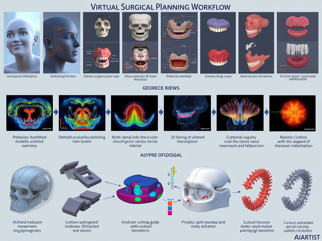
Virtual Surgical Planning (VSP)
The chapter provides a step-by-step approach to VSP:
- Preparation: Gathering and reviewing all data before the VSP session.
- Occlusion and Centric Relation Confirmation: Ensuring proper jaw relationships during planning.
- Setting Midlines and Correcting Cant: Establishing facial harmony through proper alignment.
- Determining Movements Based on TSIR: Using tooth show in repose as the primary driver for maxillary positioning.
- Reducing Interferences: Planning for removal of bony interferences.
- Surgical Sequencing: Considerations for which jaw to operate on first in double-jaw cases.
- Splint Design: Determining appropriate splint configuration and features.
The authors also discuss custom guides and hardware, their advantages and limitations, and considerations for distraction osteogenesis when large movements are planned.
Chapter 5: The Day of Surgery: Preoperative Protocols
Drs. Schlam and Jenzer detail the critical preparations required before surgery begins, emphasizing that proper setup contributes significantly to surgical efficiency and outcomes.
Operating Room Preparation
The chapter outlines essential preparations:
- Imaging and Models Display: Setting up printed or digital images, including frontal and profile views, radiographs, and VSP plans.
- Team Communication: Conducting a formal huddle with nurses, technicians, and anesthesia providers to review the surgical plan, equipment needs, and patient-specific considerations.
- Medication Considerations: Discussion of perioperative medications, including long-acting local anesthetics, tranexamic acid for blood loss reduction, and protocols for preventing postoperative nausea and vomiting.
Patient Positioning and Headwrap
Detailed steps for patient positioning and headwrap application are provided:
- Eye Protection: Using Tegaderm to protect the eyes.
- Tube Management: Recommendations for securing the nasal endotracheal tube.
- Headwrap Technique: Step-by-step instructions for creating a stable, pressure-free headwrap that allows surgical access while protecting the patient.
- Draping: Techniques for maintaining a sterile field while preserving access to facial landmarks.
The chapter concludes with a final time-out process to verify all preparations are complete before incision.
Chapter 6: Maxillary Surgery
This comprehensive chapter, led by Dr. Jenzer with contributions from Drs. Daniels, Koehler, and Arnold, provides a detailed walkthrough of various maxillary surgical procedures.
Single-Piece Le Fort I Osteotomy
The chapter provides a step-by-step approach:
- Incision and Exposure: Making a circumvestibular incision 5mm above the mucogingival junction and carefully dissecting to expose the maxilla.
- Dissection: Techniques for subperiosteal dissection to expose the maxilla, infraorbital neurovascular bundles, nasal aperture, and pterygomaxillary junction.
- Osteotomies: Detailed description of horizontal osteotomies, separation of the nasal septum, pterygoid plates, and lateral nasal walls.
- Downfracture: Digital manipulation techniques and the use of Turvey spreaders to achieve downfracture.
- Post-Downfracture Steps: Management of the descending palatine arteries, bone reduction, and preparation for fixation.
- Nasal Complex Management: Evaluation and surgical modifications of the nasal septum and inferior turbinates.
- Fixation: Techniques for proper plating and considerations for bone grafting.
- Closure: Methods for alar cinch suture placement and V-Y closure to maintain lip esthetics.
Multipiece Le Fort Procedures
The chapter extends to variations of the Le Fort osteotomy:
- Two-Piece and Three-Piece Le Fort: Techniques for interdental osteotomies, segmentation, and management of the expanded segments.
- SARPE (Surgically Assisted Rapid Palatal Expansion): Indications and techniques for expansion using distraction osteogenesis.
Throughout the chapter, the authors emphasize critical points for avoiding complications, such as maintaining a clean subperiosteal dissection to minimize bleeding and ensuring complete osteotomies to prevent unfavorable fractures.
Chapter 7: Mandibular Surgery
Dr. Czerepak and Dr. Jenzer provide an extensive guide to mandibular orthognathic procedures, focusing primarily on the Bilateral Sagittal Split Osteotomy (BSSO).
BSSO Technique
The chapter presents a detailed step-by-step approach:
- Surgical Setup: Proper positioning, preparation, and anesthesia considerations.
- Soft Tissue Dissection: Techniques for incision, lateral and medial mandibular exposure, and identification of the lingula and inferior alveolar nerve.
- Osteotomies: Detailed description of medial, oblique ridge, and anterior osteotomies, with particular attention to angles and depths to ensure favorable splits.
- Fracture Techniques: Methods for controlled fracturing of the mandible using osteotomes and chisels.
- Fixation: Positioning and fixation of the proximal and distal segments using screws or plates.
The authors emphasize critical anatomical landmarks and provide numerous tips for achieving favorable splits while avoiding complications like bad fractures or nerve damage.
IVRO (Intraoral Vertical Ramus Osteotomy)
The chapter also covers the IVRO technique:
- Indications: When IVRO might be preferred over BSSO, particularly for setbacks or cases where nerve injury must be minimized.
- Technique: Steps for performing the IVRO with attention to proximal segment positioning.
- Fixation Options: Discussion of rigid fixation versus non-rigid healing protocols.
Throughout the chapter, the authors highlight the importance of careful planning, meticulous technique, and an understanding of mandibular anatomy to achieve predictable results.
Chapter 8: Genioplasty
Drs. Ivory and Shaikh focus on chin surgery as both a stand-alone procedure and as a complement to orthognathic surgery.
Assessment and Planning
The chapter begins with methods for chin assessment:
- Clinical Evaluation: Techniques for assessing chin position, projection, and symmetry.
- Radiographic Analysis: Using lateral cephalometric radiographs and the Holdaway ratio to assess ideal chin position.
Surgical Techniques
Detailed steps for various genioplasty approaches are provided:
- Horizontal Advancement Genioplasty: Standard technique for increasing chin projection.
- Vertical Reduction/Augmentation: Methods for altering chin height.
- Asymmetry Correction: Techniques for addressing chin deviation.
- Alloplastic Augmentation: Alternative approaches using implants.
The authors emphasize the importance of proper soft tissue management, particularly of the mentalis muscle, to prevent postoperative esthetic complications.
Chapter 9: Maxillofacial Cosmetics
Dr. Serra discusses adjunctive cosmetic procedures that can enhance orthognathic surgery results.
Facial Analysis and Aging
The chapter begins with a framework for facial analysis and understanding the aging process.
Procedures
Various procedures are described:
- Rhinoplasty: Considerations when combining with orthognathic surgery.
- Lipofilling and Fat Grafting: Techniques for facial volumization.
- Submental Liposuction: Methods for improving neck contour.
- Facial Implants: Options for augmenting specific facial areas.
The chapter emphasizes the importance of understanding facial harmony and proportion when planning these adjunctive procedures.
Chapter 10: Combined Temporomandibular Joint Replacement and Orthognathic Surgery
Dr. Movahed addresses the complex challenge of managing patients with TMJ disorders who also require orthognathic surgery.
Patient Selection and Evaluation
The chapter outlines a comprehensive approach to evaluating patients for combined procedures.
Surgical Technique
Detailed steps for the following are provided:
- Total Joint Replacement: Patient-specific versus stock prosthesis considerations.
- Integration with Orthognathic Procedures: Sequencing and planning combined approaches.
- Postoperative Management: Special considerations for recovery after combined procedures.
The author emphasizes the importance of thorough planning and the benefits of addressing both issues simultaneously in appropriate cases.
Chapter 11: Maxillomandibular Advancement for Obstructive Sleep Apnea
Drs. Ivory and Figueroa focus on surgical management of obstructive sleep apnea (OSA) through orthognathic approaches.
Patient Evaluation
The chapter begins with a comprehensive approach to evaluating OSA patients:
- Sleep Studies: Interpretation of polysomnography results.
- Airway Analysis: Radiographic and clinical assessment of the airway.
Surgical Technique
Detailed steps for maxillomandibular advancement (MMA) are provided:
- Planning Considerations: Special considerations for MMA in OSA patients versus traditional orthognathic surgery.
- Technique Modifications: Enhanced advancement distances and counterclockwise rotation for maximum airway benefit.
- Postoperative Care: Special considerations for OSA patients after surgery.
The authors emphasize the effectiveness of MMA in treating moderate to severe OSA and provide guidelines for patient selection and technique optimization.
Chapter 12: Complications
Multiple contributors address the identification, prevention, and management of complications in orthognathic surgery.
Intraoperative Complications
The chapter covers management of:
- Hemorrhage: Identification and control of various bleeding sources.
- Bad Splits: Management of unfavorable fractures during BSSO.
- Nerve Injuries: Immediate management of identified nerve damage.
Postoperative Complications
Detailed discussion of:
- Infection: Prevention and management protocols.
- Malocclusion: Identification of causes and corrective approaches.
- Relapse: Understanding stability factors and management of relapse.
- TMJ Issues: Preventing and addressing postsurgical TMJ dysfunction.
- Neurosensory Disorders: Evaluation and management of postoperative nerve deficits.
The authors provide practical approaches to managing these complications, emphasizing prevention through proper planning and technique.
What Makes This Book Valuable?
Who is this book designed for?
This book is primarily designed for:
- Oral and Maxillofacial Surgery Residents: The step-by-step approach and emphasis on learning curves make it ideal for those in training.
- Practicing Surgeons: The comprehensive coverage and practical tips provide value even to experienced surgeons.
- Orthodontists: The interdisciplinary perspective helps orthodontists understand the surgical aspects of orthognathic cases.
- Dental and Medical Students: Those considering specialization in maxillofacial surgery will gain valuable insights into the field.
What are the book’s greatest strengths?
- Practical Focus: Rather than emphasizing theory alone, the book provides actionable techniques and tips that can be immediately applied in practice.
- Visual Elements: Abundant clinical photographs, diagrams, and radiographs illustrate key concepts effectively.
- Mentor-Student Approach: The authors write in a conversational style that mimics how they would teach residents in person.
- Comprehensive Coverage: The book addresses the entire workflow from initial evaluation through postoperative care.
- Complication Management: Detailed coverage of complications and their management prepares surgeons for challenging scenarios.
- Military Experience Perspective: The authors’ military backgrounds provide unique insights into efficient workflows and management of complex cases.
Are there any limitations to be aware of?
- North American Focus: The techniques and approaches primarily reflect North American practices, which may differ from those in other regions.
- Digital Workflow Evolution: While the book covers digital planning, this rapidly evolving field may see newer techniques developed since publication.
- Advanced Subspecialty Coverage: While comprehensive for orthognathic surgery, some highly specialized topics are covered more briefly than dedicated texts might offer.
Common Questions About “Introduction to Contemporary Orthognathic Surgery”
What makes this orthognathic surgery book different from others?
This book stands out for its practical, workflow-oriented approach that bridges theoretical knowledge with clinical application. Unlike many texts that focus primarily on surgical techniques alone, it provides comprehensive coverage from initial patient evaluation through postoperative care. The authors draw from their unique military surgical experiences to emphasize efficiency and troubleshooting, making it particularly valuable for learning surgeons. The mentor-student approach feels like having experienced surgeons guide you through cases step by step.
How detailed are the surgical technique descriptions?
The surgical technique descriptions are exceptionally detailed, providing step-by-step guidance with clear rationales for each decision. For example, the BSSO chapter not only outlines each cut but explains the physics behind favorable and unfavorable splits, potential pitfalls, and real-time decision-making processes. The authors include numerous clinical photographs and diagrams that illustrate key anatomical landmarks and proper instrument positioning. They also share valuable tips derived from their experience, such as methods for identifying the lingula and protecting the inferior alveolar nerve.
Does the book cover digital planning for orthognathic surgery?
Yes, the book devotes significant attention to virtual surgical planning (VSP). Chapter 4 provides a comprehensive guide to the VSP process, from preparation through execution. The authors outline an eight-step approach that includes confirming final occlusion, setting midlines, correcting cant, determining movements based on TSIR, reducing interferences, determining surgical sequencing, and designing appropriate splints. The book also discusses the advantages and limitations of custom guides and plates, offering balanced perspectives on traditional versus digital approaches.
How does the book address interdisciplinary collaboration with orthodontists?
The book includes an entire chapter (Chapter 3) written from an orthodontist’s perspective, which is relatively unique among surgical texts. This chapter helps surgeons understand orthodontic terminology, objectives, and limitations. It emphasizes the importance of communication throughout all treatment phases and explains how orthodontic decisions impact surgical planning and outcomes. The authors provide practical advice for coordinating with orthodontists, including discussions about dental decompensation, arch coordination, and management of patients who may not have been ideally prepared for surgery.
What information does the book provide about complication management?
Chapter 12 is dedicated entirely to complications, covering both intraoperative challenges (hemorrhage, bad splits, nerve injuries) and postoperative issues (infection, malocclusion, relapse, TMJ dysfunction, neurosensory disorders). The authors provide practical approaches to managing these complications, emphasizing prevention through proper planning and technique. Throughout all surgical chapters, potential complications specific to each procedure are highlighted, with preventive strategies integrated into the technique descriptions.
Does the book cover special considerations for obstructive sleep apnea patients?
Yes, Chapter 11 focuses specifically on maxillomandibular advancement for obstructive sleep apnea (OSA). The authors discuss patient evaluation, including sleep study interpretation and airway analysis. They explain how surgical planning differs for OSA patients compared to traditional orthognathic cases, including enhanced advancement distances and counterclockwise rotation for maximum airway benefit. The chapter also covers postoperative considerations specific to this patient population.
How does the book address temporomandibular joint disorders in orthognathic patients?
Chapter 10 is devoted to combined temporomandibular joint replacement and orthognathic surgery. It covers patient selection, evaluation, and detailed surgical techniques for addressing both TMJ disorders and dentofacial deformities simultaneously. The authors discuss considerations for patient-specific versus stock prostheses and provide guidance on sequencing and planning these complex procedures. Throughout other chapters, TMJ considerations are integrated into the discussion of standard orthognathic procedures.
What information is provided about patient evaluation and preparation?
The first two chapters provide extensive coverage of patient evaluation. Chapter 1 details the clinical examination process, including critical measurements like tooth show in repose, midline assessment, and cant evaluation. It also covers standardized photography protocols and provides a sample evaluation template. Chapter 2 focuses on analyzing the collected data to formulate precise diagnoses. The authors emphasize the importance of thorough preoperative preparation, including team communication and operating room setup, in Chapter 5.
Does the book cover genioplasty techniques?
Yes, Chapter 8 is dedicated to genioplasty procedures. It covers assessment methods, including the Holdaway ratio for determining ideal chin position. The authors detail various surgical approaches, including horizontal advancement, vertical modification, asymmetry correction, and alloplastic augmentation. They emphasize proper soft tissue management, particularly of the mentalis muscle, to prevent postoperative esthetic complications.
How does the book address perioperative care?
Perioperative care is covered throughout the text, with Chapter 5 specifically addressing preoperative protocols. The authors discuss medication considerations, including long-acting local anesthetics, tranexamic acid for blood loss reduction, and protocols for preventing postoperative nausea and vomiting. Postoperative care is integrated into each surgical chapter, with specific guidance on recovery, diet, oral hygiene, and follow-up schedules. The complication chapter also addresses postoperative management of various issues that might arise.
Does the book cover multipiece Le Fort osteotomies and segmental procedures?
Yes, Chapter 6 provides detailed coverage of multipiece Le Fort procedures, including two-piece and three-piece Le Forts. The authors describe techniques for interdental osteotomies, segmentation, and management of expanded segments. They discuss the critical limitation of palatal mucosa elasticity and provide practical guidelines for determining appropriate expansion distances. The chapter also covers surgically assisted rapid palatal expansion (SARPE) for cases requiring greater transverse correction.
What information is provided about fixation methods?
The book covers various fixation approaches throughout the surgical chapters. For the BSSO, the authors discuss both bicortical screws and plate fixation, with guidance on selecting appropriate methods based on case specifics. For maxillary procedures, they detail plating techniques and considerations for bone grafting. The authors also address special fixation considerations for segmental cases and asymmetry corrections. They provide balanced perspectives on traditional versus newer fixation approaches, including custom hardware.
How does the book address facial esthetics in orthognathic planning?
Facial esthetics is a core consideration throughout the text. The evaluation chapters emphasize comprehensive facial analysis, including assessment of facial thirds, profile type, and soft tissue relationships. The planning chapter highlights how tooth show in repose (TSIR) drives maxillary positioning decisions. Chapter 9 specifically addresses adjunctive cosmetic procedures that can enhance orthognathic results. Throughout the surgical chapters, the authors emphasize techniques to optimize esthetic outcomes, such as alar cinch sutures and V-Y closures to maintain lip esthetics.
Does the book cover recent advances in orthognathic surgery?
The book incorporates many contemporary approaches, including virtual surgical planning, custom guides and hardware, and digitally-driven workflows. It discusses the evolution from traditional model surgery to digital planning while maintaining balanced perspectives on both approaches. The authors address emerging topics like the use of piezoelectric devices, maxillomandibular advancement for OSA, and combined TMJ/orthognathic approaches.
What level of experience is needed to understand this book?
The book is designed to be accessible to readers at various experience levels. Residents and beginning learners will appreciate the step-by-step approach and clear explanations of fundamental concepts. More experienced surgeons will find value in the practical tips, complication management strategies, and discussions of complex cases. The authors assume basic knowledge of head and neck anatomy and surgical principles, but they explain specialized orthognathic concepts thoroughly.
Does the book address management of asymmetry cases?
Learn more about dental books
Yes, asymmetry correction is addressed throughout multiple chapters. In the evaluation and planning sections, the authors discuss methods for assessing and documenting facial asymmetry. The surgical chapters include specific considerations for asymmetry correction in both maxillary and mandibular procedures. The genioplasty chapter also addresses chin asymmetry correction. Throughout, the authors emphasize the importance of three-dimensional planning and the benefits of virtual surgical planning for complex asymmetry cases.
Conclusion: A Must-Have Resource for Orthognathic Surgery
“Introduction to Contemporary Orthognathic Surgery” represents an outstanding contribution to the field of maxillofacial surgery education. By combining comprehensive theoretical knowledge with practical clinical guidance, the authors have created a resource that will benefit clinicians at all stages of their careers.
What makes this book particularly valuable is its emphasis on the entire patient journey—from initial evaluation through final outcome. The authors don’t simply tell you what to do; they explain why and how, providing the reasoning behind each decision. This approach fosters not just technical competence but genuine understanding of orthognathic principles.
For residents and new practitioners, this book offers a structured approach to learning complex procedures. For experienced surgeons, it provides fresh perspectives and advanced techniques to enhance their practice. For orthodontists and other dental specialists, it offers valuable insights into the surgical aspects of interdisciplinary care.
In the rapidly evolving field of orthognathic surgery, with its increasing integration of digital technologies and patient-specific approaches, this text serves as both a foundational reference and a guide to contemporary innovations. The authors’ military background brings a unique perspective on efficiency, preparation, and complication management that enriches the traditional academic approach to surgical education.
Whether you’re looking to master the basics of orthognathic evaluation, refine your surgical technique, or explore advanced procedures like combined TMJ replacement, this book delivers practical, clinically relevant guidance that you can immediately apply in practice.
View more on Google