
Introduction
The Perio Handbook by Dr. Reena Wadia represents a significant contribution to the field of periodontics, offering dental professionals a thorough yet accessible guide to modern periodontal practice. As a dental student who recently completed this text, I found it combines clinical expertise with practical applications in a way that bridges the gap between theoretical knowledge and chairside techniques.
This book stands out for its systematic approach to periodontics, starting with fundamental concepts before progressing to complex treatment modalities. Dr. Wadia’s experience as the founder of RW Perio, a leading periodontal clinic in the UK, and her role as an educator through Perio School, infuses the text with evidence-based protocols and real-world applications that dental professionals at various career stages will find valuable.
In this review, I’ll provide a comprehensive summary of the book’s content, examine its strengths and limitations, and discuss which readers would benefit most from this resource. Whether you’re a dental student, general practitioner, or specialist seeking to update your periodontal knowledge, this review will help you determine if “The Perio Handbook” deserves a place in your professional library.
View more on Google
Complete Book Summary by Chapter
Part One: The Foundations
Chapter 1: The Periodontium and Beyond
What is covered in the first chapter of The Perio Handbook?
The opening chapter lays the groundwork for understanding periodontal health and disease by providing an in-depth exploration of the periodontium’s anatomy. Dr. Wadia meticulously describes the components of the periodontium, including the gingiva (both macroscopic and microscopic perspectives), periodontal ligament, root cementum, and alveolar bone.
The chapter explains that the periodontium’s main purpose is to secure the tooth to the jawbone and preserve the integrity of the masticatory mucosa’s surface. It distinguishes between the free gingiva and attached gingiva, noting their different characteristics and functions. The junctional epithelium and its role in providing attachment are highlighted as crucial elements.
The text then transitions to the epidemiology and pathogenesis of periodontitis, describing it as the most prevalent chronic inflammatory disease in humans. Dr. Wadia notes that eight out of ten people over thirty-five experience some form of gum issues, with severe periodontitis affecting 11.2% of the global population, making it the sixth most common health condition worldwide.
A significant portion of this chapter is dedicated to explaining the link between periodontitis and systemic diseases. The book explores various conditions with established connections to periodontal health, including:
- Diabetes: The book explains the bidirectional relationship, noting that diabetic patients with uncontrolled serum glucose levels are more likely to suffer from periodontitis, while periodontitis itself impacts diabetes control and increases the risk of complications.
- Cardiovascular disease: Both conditions share characteristics and mechanisms, with evidence showing they are significantly associated independent of cofounders.
- Inflammatory bowel disease: The chapter highlights the similar pathogenesis characterized by hyperactive immune responses to commensal microbiota.
- Rheumatoid arthritis: While the directionality isn’t fully established, there appears to be a clear epidemiological relationship between these conditions.
- Respiratory diseases, adverse pregnancy outcomes, fertility, neurodegenerative diseases, stress, and depression: The chapter briefly outlines the current understanding of these relationships, acknowledging where evidence is limited or emerging.
Chapter 2: Think Like a Detective
How does Dr. Wadia approach periodontal diagnosis?
Chapter 2 frames periodontal diagnosis through an investigative lens, encouraging practitioners to “think like a detective.” The chapter introduces the concept of a patient’s “causal pie,” explaining that while biofilm/suboptimal oral hygiene is always a factor, other contributing elements must be identified for accurate diagnosis and successful treatment.
The chapter outlines four quick trigger questions that serve as diagnostic clues:
- Do your gums bleed on brushing?
- Are any of your teeth loose?
- Any gum swellings or gum boils?
- Do you suffer from bad breath or notice a bad taste?
These questions not only aid diagnosis but may reveal the patient’s “pain point” – their primary motivation for seeking treatment.
Dr. Wadia then delves into systemic risk factors, defining them as occurrences or characteristics associated with increased rates of subsequent disease. The text compares biofilm to a key turning in a car’s ignition, with other risk factors “pressing on the accelerator” to drive disease progression.
The chapter covers medical history risk factors in detail:
Diabetes: The book explains that patients with hyperglycemia are three times more likely to develop periodontitis and provides practical tips for managing these patients, including ensuring they eat before appointments, checking glycemic control, and emphasizing the importance of good control.
Pregnancy: Hormonal changes magnify inflammatory effects when dental biofilm isn’t adequately controlled. The chapter offers practical guidelines for treating pregnant patients based on European Federation of Periodontology recommendations.
Immunodeficiency: The text warns clinicians to watch for necrotizing periodontal disease in immunocompromised patients, describing classic signs like pseudomembrane formation and punched-out papillae.
Medications: Certain drugs like phenytoin, cyclosporin, and calcium channel blockers can increase the risk of gingival enlargement. The chapter includes a template letter for physicians requesting medication changes when necessary.
The chapter also examines family history/genetics (noting that 50% of periodontitis susceptibility is due to host genetic factors), smoking (associated with 42% of periodontitis cases), vaping, stress, diet, and nutrition. Local risk factors like calculus, restorations with overhangs, and anatomical considerations are covered as biofilm-retentive factors.
Part Two: Assessment and Diagnosis
Chapter 3: Clinical Examination
What are the key components of periodontal clinical examination?
Chapter 3 provides a comprehensive guide to conducting a thorough periodontal examination. The chapter emphasizes that before developing a treatment plan, clinicians must perform a meticulous clinical examination to assess the periodontium’s condition.
The text begins by explaining the importance of evaluating the gingiva’s appearance across three categories:
- Color: Healthy gingivae appear “coral pink” versus inflamed tissues which appear redder
- Contour: A healthy gingiva has a scalloped shape tightly adapted to underlying tissues
- Consistency: Inflamed gingivae appear swollen and “spongy” in texture
The chapter introduces methods for objectively assessing biofilm control, describing scoring systems like the Simplified Plaque Index and the Plaque Control Record. It notes that marginal bleeding may be a more reliable measure of a patient’s ability to remove accessible plaque than plaque scores alone.
Dr. Wadia provides valuable guidance on making probing comfortable for patients through both communication and technique:
Communication tips include explaining the procedure beforehand, praising patients during the process, and using any discomfort to educate about the disease condition.
Technique tips include sweeping/dragging the probe rather than repeatedly inserting it, using appropriate pressure (approximately 25g), maintaining a finger rest, angling the probe properly along the root surface, and considering topical anesthesia when needed.
The chapter thoroughly explains the Basic Periodontal Examination (BPE), describing it as the primary and minimum standard for periodontal assessment in the UK. It outlines the three-step process for performing the BPE and explains the modified approach for patients under eighteen.
Dr. Wadia then details the six-point pocket chart, which records measurements at six sites per tooth. She explains that this more comprehensive assessment is essential for monitoring treatment response since the BPE alone cannot track how individual sites change after treatment.
The chapter concludes with guidance on assessing dental implants, noting that the BPE should not be used around implants. Instead, clinicians should complete a four- or six-point pocket chart and document the presence or absence of bleeding or suppuration on probing.
Chapter 4: Special Investigations
What special investigations are important for periodontal assessment?
Chapter 4 focuses on the additional diagnostic tools beyond clinical examination that help complete the assessment phase. The chapter is concise but emphasizes the critical role of radiographs in assessing bone levels.
Dr. Wadia explains that the type of radiograph required depends on clinical judgment and the patient’s overall dental needs. For periodontal assessment, the crestal bone levels must be visible in all areas. While bitewing radiographs may suffice for mild bone loss, periapical radiographs are often essential for accurate assessment of bone loss as a percentage of root length and visualization of periapical tissues.
The chapter provides a structured approach to radiographic interpretation, recommending that clinicians report on five key features:
- Amount of bone loss (in millimeters or percentage)
- Pattern of bone loss (horizontal or vertical)
- Local factors (evidence of calculus, restorative overhangs, or open margins)
- Anatomy (root length and morphology)
- Widening of the periodontal ligament space and signs of periapical pathology
The text emphasizes the importance of scanning radiographs in the same direction every time (either coronal to apical or apical to coronal) to ensure no important details are missed.
The chapter briefly addresses sensibility testing, explaining its value in identifying endo-perio lesions in cases of extensive disease. It mentions thermal tests (like Endo-Frost) and electric tests as methods to indirectly determine pulpal health by assessing the condition of the nerves within the dental pulp.
Chapter 5: Diagnosis
How does the book approach periodontal diagnosis and classification?
Chapter 5 demonstrates how to integrate information gathered during assessment to formulate a periodontal diagnosis. Dr. Wadia introduces the 2017 World Workshop’s classification system for periodontal and peri-implant diseases and conditions, which replaced the 1999 International Classification.
The chapter systematically covers:
Periodontal health and gingival diseases:
- Clinical health is defined as probing depths of 3mm or below with less than 10% bleeding on probing
- Gingivitis (both dental biofilm-induced and non-dental biofilm-induced) is defined and differentiated
Periodontitis:
The book provides a six-step process for diagnosing periodontitis:
- Determining if the patient has periodontitis by checking for bone loss
- Assessing the extent (localized, generalized, or molar-incisor pattern)
- Determining the stage (I-IV) based on severity
- Assigning a grade (A-C) based on progression rate
- Evaluating current disease status (stable, in remission, or unstable)
- Noting relevant risk factors (diabetes, smoking)
Necrotizing periodontal diseases:
- Characterized as among the most severe inflammatory conditions associated with dental biofilm
- Includes necrotizing gingivitis (limited to gum tissues) and necrotizing periodontitis (extends into periodontal ligament and alveolar bone)
Periodontitis as a manifestation of systemic disease:
- Cases where plaque-induced periodontitis is a key feature of a systemic disease
Other conditions affecting the periodontium:
- Systemic diseases affecting periodontal tissues
- Periodontal abscesses and endo-perio lesions
- Mucogingival deformities and conditions
- Traumatic occlusal forces
- Tooth- and prosthesis-related factors
Peri-implant diseases and conditions:
- Peri-implant health
- Peri-implant mucositis
- Peri-implantitis
The chapter provides clear diagnostic criteria for each condition and explains how to communicate these diagnoses to patients effectively.
Chapter 6: Prognosis
How should practitioners determine periodontal prognosis?
Chapter 6 discusses the importance of prognostic setting following diagnosis, explaining that it involves predicting the trajectory and outcome of a disease as well as the likelihood of recovery. The chapter emphasizes the medico-legal importance of informing patients about prognoses to prevent claims from patients who weren’t warned about teeth at risk of future loss.
Dr. Wadia outlines factors to consider when determining a tooth’s prognosis, divided into patient factors and local factors:
Patient factors include:
- Risk factors (diabetes, smoking, stress, nutrition)
- Compliance with oral hygiene and maintenance
Local factors include:
- Probing depth and tooth position
- Furcation involvement
- Amount of bone loss
- Anatomical defects
- Other associated pathology
The chapter mentions that while there is no universally accepted prognostic system, common terminology includes categories like good, fair, guarded, and hopeless, or a simplified approach using favorable, uncertain, and unfavorable. Dr. Wadia explains each category:
- Favorable: No evidence of disease or mild disease with no major risk factors or local factors, optimal oral hygiene, and good compliance
- Uncertain: Moderate level of disease with possible risk or local factors
- Unfavorable: Teeth unlikely to respond to treatment that will need extraction now or in the future
The chapter notes that prognosis may be assigned tooth-by-tooth or by grouping similar teeth, and that it may be adjusted after initial treatment once the clinician has a better understanding of the patient’s healing potential, compliance, and susceptibility.
Part Three: The Management
Chapter 7: Treatment Planning
What treatment planning approach does Dr. Wadia recommend?
Chapter 7 presents the latest evidence-based guidelines for periodontal treatment planning, referencing the European Federation of Periodontology’s S3 guidelines and the British Society of Periodontology’s implementation paper for UK practice.
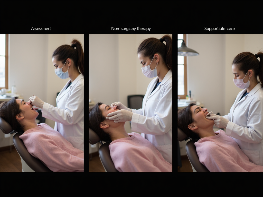
The chapter outlines a stepwise approach to treatment with four key steps:
Step 1: Supragingival PMPR and risk factor control
- Focuses on establishing oral hygiene instruction and behavioral changes
- Manages modifiable risk factors
- Includes supragingival professional mechanical plaque removal (PMPR)
- Patient “engagement” is defined as achieving ≥50% improvement in plaque and marginal bleeding scores, or plaque levels ≤20% and bleeding levels ≤30%
Step 2: Subgingival PMPR
- Implemented once the patient is engaged
- Can be performed with hand or powered instruments
- Approached via quadrant-wise or full-mouth delivery
- May include adjuncts in specific cases, though lasers and photodynamic therapy are not recommended
Step 3: Addressing residual disease
- For patients who remain unstable after Step 2
- May include non-surgical and surgical procedures
- Typically requires referral to a complexity level 2 or 3 clinician for advanced cases
Step 4: Supportive periodontal care (SPC)
- Essential for preserving periodontal stability
- Should be scheduled at intervals from three to twelve months based on risk
- May incorporate preventive and therapeutic measures from Steps 1 and 2
The chapter provides a template for a typical periodontitis treatment plan, including discussion of diabetes control or smoking cessation if relevant, oral hygiene instructions, full six-point pocket charting, supragingival PMPR, subgingival PMPR for probing depths ≥4mm with bleeding, reassessment, and lifelong supportive care.
Dr. Wadia emphasizes the importance of team-based care, with clear roles for different team members, and provides guidance on when referral to specialists is appropriate based on the British Society of Periodontology’s guidelines for patient referrals.
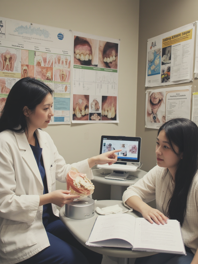
Chapter 8: Educating and Empowering Patients
How should dental professionals effectively educate and empower periodontal patients?
Chapter 8 focuses on patient education as a crucial component of successful periodontal treatment. Dr. Wadia emphasizes that the patient is at the center of the treatment team and must understand their condition and take responsibility for home care.
The chapter first addresses how to effectively communicate clinical findings to patients:
- Pockets and bleeding: Explaining pockets as spaces between tooth and gum that open and deepen with disease progression, and emphasizing that bleeding is not normal
- Radiographic findings: Using radiographs to show bone loss and demonstrate the damage caused by periodontitis
- Diagnosis: Ensuring patients understand their periodontal diagnosis and its severity
- Treatment plan: Translating the treatment plan to focus on what matters to the patient, such as keeping teeth longer rather than technical outcomes like “improved probing depths”
Dr. Wadia then outlines a five-step process for effective home care instruction:
- Orientate: Introduce basic oral anatomy using mirrors or intraoral cameras
- Disclose: Use disclosing agents to highlight missed areas during brushing
- Demonstrate: Have patients show their current technique and then adapt as needed
- Reinforce responsibility: Emphasize that 80% of treatment success depends on home care
- GPS (Goal setting, Planning, Self-monitoring): An evidence-based technique for behavioral change
The chapter discusses behavioral change models, including Oral Hygiene TIPPS and the COM-B model, which emphasizes capability, opportunity, and motivation as essential for behavior change.
Dr. Wadia provides specific recommendations for oral hygiene tools:
- Power brushes over manual toothbrushes
- Interdental brushes as the first choice for interdental cleaning
- Single-tufted brushes for hard-to-reach areas
- Tongue cleaning for patients with tongue coating
The chapter includes creative analogies to help explain oral hygiene concepts to patients, such as comparing toothpaste to face cream and interdental brushes to essential medication. It concludes by addressing biofilm-retentive factors, including anatomical factors and iatrogenic factors that can complicate oral hygiene.
Chapter 9: Non-Surgical Periodontal Therapy
What does effective non-surgical periodontal therapy involve?
Chapter 9 delves into the practical aspects of non-surgical periodontal therapy, emphasizing that the aim is to “close” pockets (achieve probing depths of 4mm without bleeding or below) through a combination of gingival recession, long-junctional epithelium formation, resolution of inflammation, and cuff tightening.
The chapter outlines expected improvements from non-surgical therapy based on research:
- For shallow sites (≤5mm): mean reduction of 1.5-1.6mm
- For deep sites (≥7mm): mean reduction of 2.6mm
- Pocket closure is most predictable around anterior teeth and least likely around molars
Dr. Wadia emphasizes the importance of obtaining informed consent, discussing four key areas with patients:
- Pain (manageable with standard analgesics)
- Bleeding (may initially increase before settling)
- Sensitivity (typically short-term)
- Gum recession (a sign of healing as swelling reduces)
The chapter provides detailed guidance on local anesthesia techniques to ensure patient comfort, including creating a calming environment, using topical anesthesia, employing pressure distraction, and ensuring slow deposition of solution.
Dr. Wadia discusses the choice between quadrant, half-mouth, or full-mouth approaches, noting that clinical outcomes are generally similar. The decision should be based on disease severity, time availability, anesthesia use, patient anxiety, biofilm score, and medical considerations.
The text addresses adjuncts to non-surgical therapy:
- Chlorhexidine (CHX) mouthwash for limited time in specific cases
- Antibiotics in limited situations, with azithromycin being a popular choice due to once-daily dosing
The chapter introduces the concept of Minimally Invasive Non-Surgical Therapy (MINST), a novel approach focused on biofilm removal without intentional loss of tooth structure, particularly beneficial for intrabony defects.
Dr. Wadia provides comprehensive guidance on instrumentation techniques:
- Ultrasonics: Explains mechanisms of action, proper technique, and tips for effective use
- Hand instrumentation: Details the parts of hand instruments and how to select and use them appropriately
- Air polishing: Describes the benefits of AIRFLOW® with PLUS® powder
The chapter concludes with advice on preventing repetitive strain injury (RSI) and back/neck pain during periodontal therapy, encouraging proper positioning and the use of magnification.
Chapter 10: Periodontal Challenges
How should clinicians manage common periodontal challenges?
Chapter 10 addresses specific challenges encountered in periodontal management, beginning with occlusal trauma and splinting. Dr. Wadia defines traumatic occlusal force as “any occlusal force resulting in injury to the teeth and/or the periodontal attachment apparatus,” and distinguishes between primary occlusal trauma (affecting teeth with healthy support) and secondary occlusal trauma (affecting teeth with weakened support).
The chapter outlines indications for permanent splinting:
- Immobilizing mobile teeth causing discomfort or affecting function
- Immobilizing teeth with progressively increasing mobility
- Preventing further tooth movement
Dr. Wadia emphasizes that periodontitis must be addressed before splinting and recommends archwire and composite when possible, as this allows some independent tooth movement.
A significant portion of the chapter focuses on furcation management. Dr. Wadia defines furcation defects as “bone resorption and interradicular attachment loss in multirooted teeth” and notes that maxillary molars are the most difficult to assess and treat due to multiple furcation entrances in proximal areas.
The chapter presents a comprehensive overview of furcation management options:
- Non-surgical management (for Grade I furcations with good access)
- Surgical approaches (resective or regenerative)
- Furcation-plasty (odontoplasty and osteoplasty)
- Tunnel preparation (for molars with short root trunks and divergent roots)
- Root resection (removing one or two roots while retaining the crown)
- Hemisection (removing a root and part of the crown it supports)
- Extraction when other options aren’t viable
The text then addresses dentine hypersensitivity (DH), a common issue in periodontitis patients, especially following treatment. Dr. Wadia explains Brannstrom’s hydrodynamic theory and outlines a management approach starting with modifying etiological factors, then recommending over-the-counter products, and finally applying professional agents for severe cases.
The chapter concludes with a detailed discussion of halitosis (bad breath), affecting approximately 30% of the population. Dr. Wadia explains that 85% of persistent genuine halitosis originates from inside the mouth, primarily from tongue coating, gingivitis, and periodontitis. The recommended management approach includes personalized guidance, smoking cessation, maintaining oral moisture, optimizing oral hygiene, implementing tongue cleaning, suggesting chemical agents when necessary, providing periodontal treatment, and addressing local dental factors.
Chapter 11: Periodontal Emergencies and Their Management
How should periodontal emergencies be managed?
Chapter 11 provides a comprehensive guide to recognizing and managing various periodontal emergencies, beginning with gingival abscesses. Dr. Wadia defines these as “localised purulent infections affecting the marginal gingiva or interdental papillae,” typically presenting as rapidly expanding localized swellings that may be shiny, smooth, or pointed.
The management approach for gingival abscesses includes incision, drainage, irrigation with saline, addressing the underlying cause, and potentially recommending chlorhexidine mouthwash or warm saline rinses.
The chapter then addresses periodontal abscesses, defined as localized pus collections within the gingival wall of a periodontal pocket. These present as ovoid swellings on the lateral side of the tooth root, typically associated with deep periodontal pockets. Dr. Wadia outlines various causes, including closure of pocket margins in complex morphology, furcation involvement, or vertical defects.
Management of periodontal abscesses focuses on establishing drainage through the periodontal pocket or external incision, cleaning the area, and adjusting occlusion for immediate relief. Antibiotics are recommended only for systemic involvement or spreading infection.
The text covers pericoronitis/peri-coronal abscesses, which affect partially erupted teeth (most commonly mandibular third molars). Management involves irrigating the operculum to remove debris, potentially excising the operculum, and adjusting the opposing tooth to prevent trauma.
Dr. Wadia thoroughly discusses necrotizing gingivitis and periodontitis, characterized by necrosis and ulcers in the free gingiva with a “punched-out” appearance of the interdental papilla. Treatment involves careful superficial debridement, preferably with ultrasonics, daily debridement during the acute phase, and chemical plaque control with chlorhexidine. Systemic antimicrobials like metronidazole may be necessary if debridement alone is insufficient.
The chapter also addresses acute herpetic gingivostomatitis, a viral infection presenting as small ulcers with raised edges across various oral mucosal surfaces. Since the condition is self-limiting, treatment focuses on hydration and pain management.
Dr. Wadia discusses physical, chemical, and thermal injuries to periodontal tissues, noting that proper diagnosis requires thorough clinical examination and patient history to identify the cause. Treatment involves eliminating the initiating factor and addressing pain symptoms.
The chapter concludes with subgingival root fractures, which can cause acute pain and lead to periodontal infection. Diagnosis may require magnification and good lighting, with fracture lines not always visible. Management depends on the tooth’s vitality and the fracture’s location and extent, potentially including endodontic therapy, full coverage restoration, periodontal flap for visualization, or extraction if untreatable.
Chapter 12: Supportive Periodontal Care
Why is supportive periodontal care essential and how should it be implemented?
Chapter 12 emphasizes the critical importance of supportive periodontal care (SPC) in maintaining treatment outcomes, stating that with proper SPC, periodontal health can be maintained in most patients, even in advanced cases.
The chapter outlines three key aims of SPC:
- Monitoring periodontal stability and initiating treatment if progression is detected
- Removing etiological factors before disease progresses
- Reinforcing smoking cessation and organizing referral if appropriate
Dr. Wadia provides detailed guidance on determining recall intervals based on the patient’s risk profile. The default interval is three months but can extend up to twelve months depending on various factors:
Patient factors:
- Smoking status (smokers require more frequent appointments)
- Systemic conditions like uncontrolled diabetes
- Stress levels
- Genetic markers like IL-1 genotype positivity
- Previous susceptibility to severe periodontitis (Grade C)
Previous treatment factors:
- Type of treatment (surgical cases may need more frequent monitoring)
- Treatment outcome limitations
Local factors:
- Biofilm management capability
- Type of periodontal disease (infrabony lesions, furcation involvement)
- Severity of past or existing disease
- Concurrent oral conditions
The chapter mentions risk assessment tools like PreViser/DEPPA and the Periodontal Risk Assessment (PRA) “spider diagram” that can help determine appropriate recall intervals.
Dr. Wadia introduces the concept of palliative periodontal care for cases where a patient enters SPC with acceptance of deep pockets. In these situations, the aim is to slow disease progression rather than improve the condition, typically when risk factors remain unchanged (e.g., continued smoking) or after multiple rounds of active treatment without improvement.
The chapter concludes with a structured approach to SPC appointments, which typically include:
- Medical history review
- Oral hygiene regime review
- Risk factor control assessment
- Comprehensive probing
- Supragingival PMPR as needed
For partially successful non-surgical cases with residual deep pockets, appointments would also include subgingival PMPR of these sites. The chapter notes that significant relapse may occasionally necessitate re-entering the active treatment phase before returning to SPC.
Chapter 13: Dental Implants
What considerations are important for dental implants in periodontitis patients?
Chapter 13 begins with a brief history of dental implants, crediting Professor Brånemark as the pioneer of titanium endosseous dental implants and introducing the concept of osseointegration. Dr. Wadia emphasizes that patients must understand that implant-supported restorations require ongoing maintenance and can develop complications like peri-implantitis.
The chapter explains key terminology related to implants, including osseointegration, endosseous dental implant, implant abutment, abutment screw, and single-stage versus two-stage implant surgery.
Dr. Wadia highlights fundamental differences between natural teeth and dental implants:
- The periodontal complex around teeth has evolved over millions of years, while the tissue around implants is essentially scar tissue
- Osseointegration occurs as a reaction to foreign material
- Peri-implant tissues are more susceptible to inflammatory reactions compared to periodontal tissues
The chapter then addresses specific considerations for periodontitis patients receiving dental implants:
Risk of peri-implant disease:
- Patients with a history of periodontitis face an increased risk of developing peri-implantitis
- Long-term studies show greater probing depths, bone loss, and failure rates in these patients
- Periodontitis should be addressed prior to implant placement
- Patients should be informed of their increased risk during the consent process
Implant position:
- Implants should be spaced at least 3mm apart to facilitate biofilm management
- Improper positioning can result in difficult-to-clean restorations
- Placing implants too deeply can complicate biofilm control
Screw versus cement:
- Screw-retained restorations are recommended when possible
- Excess cement can create rough surfaces that facilitate microbial colonization
Removable options:
- Overdentures may be a more easily cleaned and safer long-term solution
- Can replace both hard and soft tissue for better aesthetics
- Allow better access to peri-implant tissues for ongoing plaque control
Supportive peri-implant care:
- Lack of supportive therapy is associated with higher frequency of peri-implantitis
- Recall intervals should be tailored to individual risk factors
Extracting teeth to replace with implants:
- Growing evidence suggests maintaining periodontally involved teeth can provide better outcomes than implant replacement
- A trend toward retaining periodontally involved teeth for as long as possible
The chapter presents key recommendations from the European Federation of Periodontology regarding dental implants, emphasizing patient awareness, risk assessment, proper component fit, tissue management, and personalized supportive care.
The chapter concludes with guidance on managing peri-implant disease, progressing from reinforcing self-care for healthy implants to professional management of peri-implant mucositis and referral for peri-implantitis cases.
Chapter 14: Multidisciplinary Care
How does periodontal care interact with other dental specialties?
Chapter 14 explores the integration of periodontal care with other specialties, focusing primarily on the perio-endo and perio-ortho interfaces.
The perio-endo interface section begins by defining endo-perio lesions as pathological communications between pulpal and periodontal tissues that can be acute or chronic. Dr. Wadia illustrates the various routes of communication, including exposed dentinal tubules, lateral and accessory canals, and the apical foramen.
Key signs of endo-perio lesions include deep periodontal pockets extending to the root apex and negative or altered response to pulp vitality tests. Additional signs may include radiographic bone loss, spontaneous pain, suppuration, mobility, sinus tracts, and color changes.
For treatment planning, Dr. Wadia notes that prognosis is more predictable when the pulp is the primary cause but becomes less certain when significant periodontal damage has impacted the root canal. The recommended approach is to perform endodontic treatment first, followed by reassessment of the periodontal condition 2-3 months later.
The chapter then transitions to the perio-ortho interface, emphasizing the challenges of orthodontics in patients with severe periodontitis. Dr. Wadia stresses the importance of achieving periodontal health (shallow, maintainable pockets and controlled inflammation) before initiating orthodontic treatment.
The text specifically addresses orthodontics in Stage IV periodontitis, characterized by anatomical and functional consequences due to tooth and periodontal attachment loss. Dr. Wadia references clinical practice guidelines that emphasize interdisciplinary collaboration for rehabilitating compromised dentition.
The chapter provides detailed information on tooth movements in periodontitis patients:
- Pathological tooth migration (drifting, flaring, elongation) can be corrected orthodontically
- Orthodontic movements like intrusion, retraction, and alignment are unlikely to negatively affect periodontal conditions
- Orthodontic treatment can improve the height of interdental papillae and potentially reduce tooth mobility
- For teeth with intrabony defects, combined orthodontic treatment can safely occur after periodontal regenerative procedures
Dr. Wadia explains the biomechanical considerations for orthodontic treatment in periodontitis patients:
- The center of resistance shifts further apically in teeth with diminished periodontal support
- Tipping movements are easier to achieve but create uneven stress distribution
- Light, controlled forces should be applied to mitigate risks of hyalinization and bone damage
The chapter compares fixed versus removable orthodontic appliances, noting that fixed appliances are generally recommended for patients with advanced periodontitis. It also discusses the value of skeletal anchorage devices for enhancing treatment effectiveness.
Dr. Wadia addresses potential complications during orthodontic treatment in periodontitis patients, including increased biofilm retention, gingival enlargement, and root resorption. She emphasizes the importance of close monitoring and pausing active treatment if periodontal issues arise.
The chapter concludes with guidance on post-orthodontic maintenance and retention, recommending permanent fixed passive retainers, potentially combined with removable retainers, with ongoing supportive measures to detect retainer failures and monitor periodontal stability.
Chapter 15: Pocket Reduction Surgery
What surgical approaches are available for pocket reduction?
Chapter 15 begins with a historical perspective on periodontal surgery, tracing its evolution from the removal of diseased tissue to a focus on pocket elimination, and finally to the current emphasis on accessing root surfaces for effective debridement and creating gingival contours that facilitate self-performed biofilm control.
Dr. Wadia outlines three main aims of pocket reduction surgery:
- Allowing access and visibility for adequate debridement
- Establishing a favorable dentogingival architecture to support oral hygiene
- Reducing probing depths and possibly restoring the periodontal apparatus when there has been attachment loss
The chapter emphasizes the importance of completing at least one, ideally two, sessions of non-surgical periodontal therapy before considering surgical intervention. This initial phase helps eliminate local risk factors and allows evaluation of the patient’s oral hygiene practices and compliance.
Dr. Wadia introduces the concept of critical probing depth as a guide for clinical decision-making, noting that flap surgery is generally indicated for probing depths exceeding 5.4mm (commonly rounded to 6mm), while non-surgical options are preferred for depths between 2.9-5.4mm.
The chapter outlines the general steps in pocket reduction surgery:
- Flap design/incisions
- Removal of excised tissues (if resective surgery)
- Raising the flap
- Debridement of root surfaces and bony defects
- Management of bone
- Checking flap apposition and adjusting if needed
- Suturing
Dr. Wadia explains that various resective techniques exist for horizontal bone loss, while regenerative approaches are increasingly used for intrabony defects. She describes three types of defects: suprabony (horizontal), intrabony (vertical), and interradicular (furcation), noting that regenerative surgery is indicated for the latter two when suitable.
The chapter provides detailed information about bone grafting materials used in regenerative procedures, including:
Enamel matrix derivative (Emdogain):
- Contains amelogenin, a hydrophobic protein that functions as a cell-adhesion matrix-bound protein
- Plays a crucial role in wound healing and soft tissue regeneration
- Affects cell attachment, proliferation, differentiation, and survival
Bio-Oss®:
- Bovine-derived natural bone substitute
- Integrates into newly formed bone matrix
- Helps maintain volume over extended periods
Bio-Gide®:
- Porcine-derived resorbable collagen membrane
- Prevents soft tissue ingrowth into the augmented site
- Guides bone, soft tissue, and blood vessel development
The chapter notes that current evidence shows true periodontal regeneration (periodontal ligament, cementum, and bone) with decalcified freeze-dried bone allograft, demineralised bovine bone mineral, and enamel matrix derivative. It concludes by mentioning the trend toward microsurgical and minimally invasive approaches that enable careful flap preparation and suturing, resulting in reduced tissue damage and more effective healing.
Chapter 16: Gingival Recession and Surgery
How should gingival recession be evaluated and managed surgically?
Chapter 16 focuses on gingival recession, defined as the apical displacement of the gingival margin resulting in exposure of the tooth root. Dr. Wadia notes this condition can significantly impact aesthetics and is a common reason patients seek periodontal treatment.
The chapter first examines the multifaceted etiology of gingival recession, distinguishing between predisposing factors (which increase risk) and precipitating factors (which trigger onset):
Predisposing factors:
- Thin gingival phenotype (visible probe through tissue during probing)
- High frenal attachments and shallow vestibular depths
- Thin buccal bone, dehiscences, or fenestrations
- Tooth positioning and root-bone mismatch
- Loss of interproximal bone support
Precipitating factors:
- Trauma (aggressive brushing, improper flossing, piercings, malocclusion)
- Plaque accumulation leading to inflammation
Dr. Wadia emphasizes the clinician’s responsibility to identify and manage modifiable factors, as unaddressed factors like aggressive brushing will undermine surgical interventions.
The chapter explains the current classification system for gingival recession (from the 2018 World Workshop), which divides recession into three types:
- RT1: No loss of interproximal attachment
- RT2: Interproximal attachment loss less than or equal to buccal attachment loss
- RT3: Interproximal attachment loss greater than buccal attachment loss
The text distinguishes between trauma-induced recession (progressing from the exterior inward) and bacterial-induced recession (progression from inside outward).
Dr. Wadia outlines a treatment approach that begins with addressing modifiable etiological factors and monitoring recession when surgery isn’t clearly indicated. Surgical intervention may be considered for aesthetics, augmenting keratinized tissue/thickness, or preventing recession progression.
The chapter describes various surgical techniques:
Pedicle soft-tissue graft procedures:
- Rotational flap procedures (laterally sliding flap, double papilla flap, oblique rotational flap)
- Advanced flap procedures (coronally advanced flap, semilunar coronally repositioned flap)
- Regenerative procedures
- Tunneling
Free soft-tissue graft procedures:
- Epithelialized graft
- Subepithelial connective tissue graft
Dr. Wadia provides detailed information about three common approaches:
Free gingival graft: Enhances keratinized tissue height, thickens gingival tissue, and deepens the vestibule, primarily to promote periodontal health rather than improve aesthetics.
Coronally advanced flap: A pedicle soft-tissue graft procedure involving a coronal shift of soft tissues over the exposed root surface, ideal for isolated recession defects.
Tunnel procedure: Leaves interdental papillae intact, with a connective tissue graft placed in the tunnel and potentially coronally positioned to allow complete coverage of the graft.
The chapter concludes by noting that tissue graft procedures work best for patients with good bone levels and interdental support, and that healing involves a mix of repair and regeneration. Results are likely to last provided etiological factors have been addressed.
Chapter 17: Crown-Lengthening Surgery
What is crown-lengthening surgery and when is it indicated?
The final chapter of Part Three focuses on crown-lengthening surgery, another type of periodontal surgical treatment that aims to resolve gingival excess for either aesthetic or functional reasons.
Dr. Wadia begins by defining crown lengthening as “the surgical removal of hard and soft periodontal tissue to gain supragingival tooth length, allowing for longer clinical crowns and the re-establishment of the biologic width (now called ‘supracrestal tissue attachment’).” This attachment encompasses the junctional epithelium and supracrestal connective tissue and should be approximately 2mm.
The chapter divides crown-lengthening procedures into two main categories:
- Aesthetic: Addressing excessive gingival display, short clinical crowns, and uneven gingival contours
- Restorative/functional: Needed when there is insufficient sound tooth structure for restoration, to access subgingival fractures or caries, or to reposition crown margins
Dr. Wadia describes ideal “pink” aesthetics, noting that a smile is generally considered pleasant when it exposes all maxillary teeth with approximately 1mm of facial gingiva. The chapter details the components of ideal gingival architecture:
- Gingival zenith position: The lowest point of gingival contour, ideally positioned 1mm distal to the midline for central incisors, 0.4mm distal for lateral incisors, and centrally along the long axis for canines
- Relative gingival margins: Lateral incisors’ zeniths should be 1mm coronal to those of centrals and canines
- Gingival aesthetic line: Connecting the tangents of central incisor and canine zeniths, ideally forming a 45-90 degree angle with the midline
The chapter explores various causes of gingival excess, including altered passive eruption, dentoalveolar extrusion due to tooth wear, vertical maxillary excess, and short or hypermobile upper lip. Dr. Wadia emphasizes the importance of proper diagnosis, as some conditions (like vertical maxillary excess) may require maxillofacial surgery rather than periodontal intervention.
The chapter outlines a three-stage treatment sequence for crown lengthening:
1. Planning:
- Establishing periodontal health and effective plaque control
- Evaluating local factors like crown-to-root ratio and root proximity
- Identifying the reference point (CEJ, existing restorations, or surgical stent)
- Assessing gingival architecture and keratinized tissue
- Determining bone position relative to the reference point
2. Surgery:
- For aesthetic cases: buccal flap elevation and exclusive buccal bone removal
- For restorative cases: both buccal and palatal/lingual flap elevation with bone removal on all surfaces
- Bone removal (ostectomy) to re-establish the supracrestal tissue attachment
- Suturing with single interrupted or mattress sutures
3. Further restorative treatment:
- For altered passive eruption, additional restorative work may not be necessary
- For other cases, provisional restorations can be placed after suture removal
- Definitive restorations in the aesthetic zone may be delayed for six months as the gingival position continues to evolve
The chapter concludes by noting that coronal displacement of the gingival margin tends to be more significant in individuals with thick gingival phenotypes, and patients should be informed about the possibility of revision surgery.
Conclusion
The final section of the book offers a concise conclusion that encapsulates Dr. Wadia’s mission in writing this comprehensive guide. She expresses hope that readers now feel equipped with a robust foundation in essential periodontal concepts and practices, enabling them to deliver exceptional care.
The conclusion emphasizes that by exploring the foundations of periodontal disease, mastering assessment and diagnosis, and learning various treatment strategies, readers should be well-prepared to address the many challenges that arise in professional practice. Dr. Wadia reiterates that her goal has been to empower dental professionals with knowledge and tools that directly translate to improved patient outcomes.
She encourages readers to use the book as an ongoing reference in their professional journey, providing support at every step from integrating new techniques to managing periodontal emergencies and planning surgical interventions.
In-Depth Review
What makes The Perio Handbook stand out from other periodontal textbooks?
The Perio Handbook distinguishes itself through its remarkably practical approach to periodontics. Unlike many academic textbooks that focus heavily on theory, Dr. Wadia consistently emphasizes chairside application and real-world scenarios. The book strikes an ideal balance between evidence-based protocols and clinical pragmatism, making it immediately useful for practitioners.
Another standout feature is the book’s accessibility. Complex concepts are explained clearly without oversimplification, making the content digestible for students and general practitioners while still offering depth for specialists. Dr. Wadia’s writing style is direct and engaging, avoiding the dense, jargon-heavy prose that characterizes many dental textbooks.
The handbook’s comprehensive yet concise nature gives it exceptional versatility. It thoroughly covers the entire spectrum of periodontal practice—from basic concepts to advanced surgical techniques—without becoming unwieldy. This makes it equally valuable as a quick reference during clinical practice, a study guide for students, or a refresher for experienced clinicians.
The emphasis on patient communication and behavioral change represents another significant strength. Dr. Wadia dedicates substantial space to discussing how to effectively educate patients, motivate compliance, and translate clinical findings into language that resonates with patients—skills often overlooked in traditional periodontal education.
What are the book’s strongest sections?
The diagnostic sections of The Perio Handbook are particularly strong. Dr. Wadia masterfully explains the 2017 classification system, breaking down the potentially confusing staging and grading approach into a clear, step-by-step process that clinicians can readily apply.
The non-surgical therapy chapters offer exceptional clinical value, providing detailed guidance on techniques, instrumentation, and expected outcomes. The inclusion of practical tips—from making probing comfortable to preventing repetitive strain injury—demonstrates Dr. Wadia’s extensive clinical experience and attention to the everyday challenges practitioners face.
The sections on patient education and behavioral change stand out for their innovative approaches. The GPS (Goal setting, Planning, Self-monitoring) framework and creative analogies for explaining oral hygiene concepts provide practitioners with immediately applicable tools for improving patient compliance.
The multidisciplinary care chapters excel at explaining the interfaces between periodontics and other specialties. The perio-ortho section, in particular, provides clarity on a complex topic, addressing biomechanical considerations specific to periodontally compromised patients that aren’t commonly discussed in either periodontal or orthodontic literature.
What areas could be improved?
While the book provides excellent clinical protocols, some sections could benefit from more case studies to illustrate the application of these protocols in different patient scenarios. Visual examples showing pre-treatment, mid-treatment, and post-treatment outcomes would enhance understanding, particularly for surgical procedures.
The emerging field of digital technologies in periodontics—including digital smile design, CBCT-guided surgery, and 3D-printed surgical guides—receives relatively limited coverage. As these technologies become more integrated into mainstream practice, future editions might expand on these topics.
Although the book references current research, some sections could provide more detailed discussion of controversial areas where evidence is evolving or conflicting. This would help readers develop critical thinking skills for navigating areas where clear consensus doesn’t yet exist.
Who would benefit most from reading this book?
General dental practitioners would find tremendous value in this handbook, as it provides clear, actionable guidance for managing the periodontal needs of their patients. The emphasis on non-surgical therapy and knowing when to refer makes it particularly relevant to this audience.
Dental students and recent graduates will appreciate the book’s systematic approach and clear explanations, which build logically from foundation concepts to complex treatments. It effectively bridges the gap between academic learning and clinical application, serving as an ideal companion throughout their educational journey.
Dental hygienists and therapists will find the sections on assessment, non-surgical therapy, and supportive care especially valuable, as these areas align closely with their scope of practice. The practical tips for effective instrumentation and patient education directly address their daily clinical challenges.
Specialists in periodontology may find the book most useful as a teaching aid or quick reference, particularly for its clear explanation of the current classification system and its practical approach to patient communication and motivation.
Conclusion
Is The Perio Handbook worth adding to your professional library?
The Perio Handbook represents an exceptional addition to any dental professional’s library. It successfully delivers on its promise to provide a comprehensive yet accessible guide to modern periodontics, covering everything from fundamental concepts to advanced surgical techniques. What sets it apart is its unwavering focus on practical application and its clear, systematic approach to periodontal care.
Dr. Wadia has created a resource that doesn’t just explain periodontal concepts but translates them into actionable protocols that can be immediately implemented in clinical practice. The book excels at addressing the everyday challenges practitioners face, from making probing comfortable for anxious patients to managing complex emergencies.
For students and general practitioners, this handbook offers a clarity and accessibility rarely found in periodontal textbooks. For specialists, it provides a valuable reference and teaching tool that reflects current evidence-based practice. For the entire dental team, it emphasizes the collaborative approach essential for successful periodontal outcomes.
While no single book can cover every aspect of a dynamic field like periodontics, The Perio Handbook comes remarkably close to being a complete resource. Its few limitations are far outweighed by its practical value and comprehensive scope. Whether you’re beginning your dental career or looking to update your periodontal knowledge, this book represents an investment that will pay dividends in improved patient care and clinical confidence.
Short description: A comprehensive review of “The Perio Handbook” by Dr. Reena Wadia, examining its content, strengths, and practical applications for dental professionals at all career stages. The article provides a detailed chapter-by-chapter summary and evaluates the book’s value for different readers, emphasizing its practical approach to modern periodontal practice.
Learn more about Dental Books